Cost: A transvaginal ultrasound (TVS) in Nepal costs between NPR 700 and NPR 3,000, depending on the facility. Government hospitals charge NPR 500–800, while private fertility clinics in Kathmandu typically charge NPR 1,500–3,000. At Sishu Fertility Clinic, the TVS scan is priced at NPR 700.
Procedure: A thin, lubricated probe is gently inserted into the vagina to produce detailed images of the uterus, ovaries, fallopian tubes, and cervix. The scan takes 15–30 minutes and requires an empty bladder beforehand. No anaesthesia or recovery time is needed.
Pain: For most women, a TVS scan is not painful. The sensation is mild pressure or a feeling of fullness, similar to a routine pelvic examination. The probe is smaller than a standard speculum used in a cervical smear test.
Safety: TVS is completely safe. It uses sound waves, not radiation, so it carries no risk of radiation exposure. The probe is covered with a sterile single-use sheath before every scan. TVS is safe during pregnancy, safe with an IUD, and safe to repeat multiple times.
This guide covers everything you need to know before your TVS scan at Sishu Fertility Clinic in Kathmandu: what it is, why it is done, exactly what happens during the procedure, how much it costs in Nepal, and how it compares to an abdominal ultrasound.
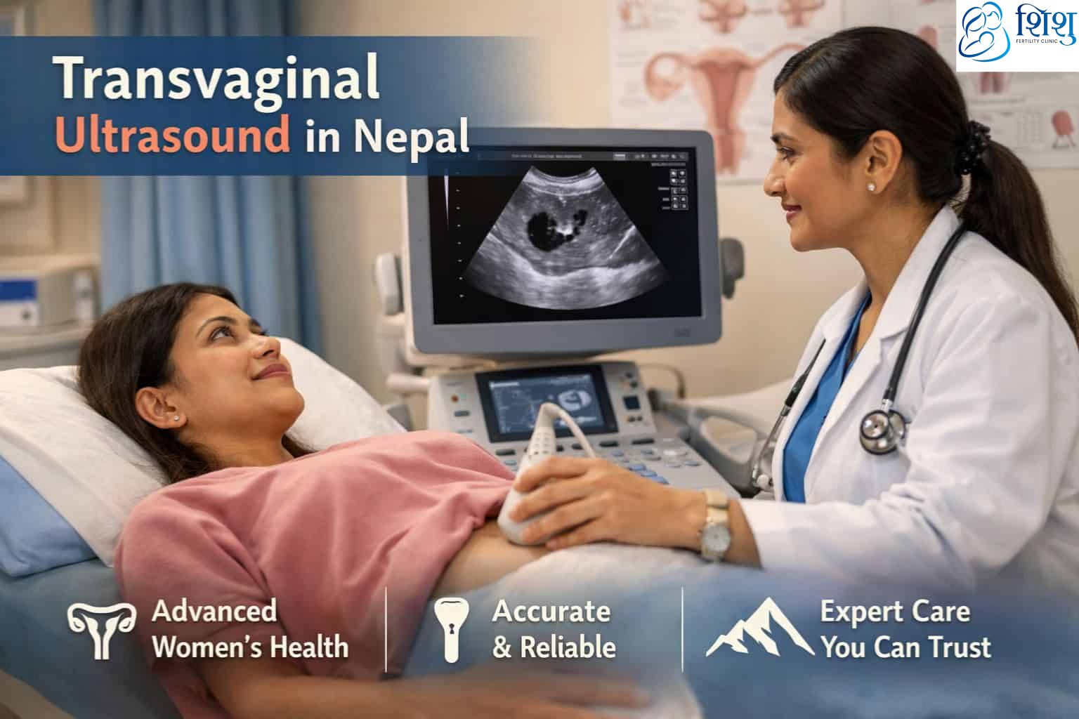
What Is TVS – Transvaginal Ultrasound?
TVS stands for Transvaginal Sonography (also written as Transvaginal Ultrasound or TVS scan). It is a type of ultrasound that uses high-frequency sound waves, not radiation, to create real-time images of the female reproductive organs.
Unlike a standard abdominal ultrasound where the probe is placed on top of the skin, a TVS scan uses a thin, wand-shaped probe (called a transducer) that is inserted a short distance into the vaginal canal. Because the probe is closer to the organs being examined, the images it produces are significantly sharper and more detailed than what an abdominal scan can achieve.
TVS is a standard, widely used diagnostic tool in obstetrics and gynaecology. According to medical literature published in the Journal of Armed Forces India, transvaginal sonography provides superior visualisation of pelvic structures compared to transabdominal scanning, particularly in women who are overweight or in early pregnancy where an abdominal scan may produce unclear images.
TVS vs Abdominal Ultrasound – What Is the Difference?
Many women are referred for a TVS scan after having had an abdominal ultrasound. Understanding the difference helps clarify why your doctor is recommending it.
| Transvaginal Ultrasound (TVS) | Abdominal Ultrasound | |
|---|---|---|
| Probe placement | Inside the vagina | On the abdomen skin surface |
| Image quality | High detail – closer to organs | Lower detail – further from organs |
| Bladder preparation | Empty bladder required | Full bladder usually required |
| Best for | Early pregnancy, ovarian cysts, IVF monitoring, uterine lining | Later pregnancy, general pelvic overview |
| Discomfort level | Mild pressure | None |
| Radiation | None | None |
The TVS scan is preferred in gynaecology and fertility settings because it gives the specialist a much clearer view of the uterine lining thickness, follicle size, ovarian cysts, and early pregnancy, all of which are difficult to assess accurately with an abdominal scan alone.
Why Is a TVS Scan Done? Common Reasons

A transvaginal ultrasound is recommended for a wide range of gynaecological and fertility-related purposes. Your doctor may request a TVS scan for any of the following reasons:
Fertility assessment and IVF monitoring
TVS is an essential part of any fertility evaluation. It is used to count antral follicles (AFC) to estimate ovarian reserve, monitor follicle growth during ovarian stimulation in an IVF cycle, and guide the egg retrieval procedure. If you are undergoing IVF treatment in Nepal, you will have multiple TVS scans throughout your cycle.
Diagnosing ovarian cysts
TVS provides clear imaging of both ovaries, allowing the specialist to identify, measure, and classify ovarian cysts. It can distinguish between simple fluid-filled cysts and more complex structures that may require further investigation.
Evaluating the uterus
TVS can detect uterine fibroids (non-cancerous growths in the uterine wall), uterine polyps (small growths on the inner lining), and structural abnormalities such as a septate uterus. The endometriosis page on this site explains how TVS is also used as part of the diagnostic workup for endometriosis — a condition where tissue similar to the uterine lining grows outside the uterus.
Early pregnancy monitoring
TVS is the most reliable way to confirm an intrauterine pregnancy in the early weeks, detect a fetal heartbeat from around 6 weeks of gestation, and identify potential complications such as ectopic pregnancy or threatened miscarriage.
Investigating abnormal uterine bleeding
Unexplained heavy periods, bleeding between periods, or post-menopausal bleeding are all indications for a TVS scan to examine the uterine lining and look for polyps, fibroids, or thickening that may need further evaluation.
Investigating pelvic pain
TVS helps identify the cause of unexplained pelvic pain, including ovarian cysts, pelvic inflammatory disease (PID), or endometriosis.
Assessing the AMH and ovarian reserve
TVS is performed alongside the AMH blood test to give a complete picture of a woman’s ovarian reserve. The antral follicle count seen on TVS complements the AMH level to help predict how a woman will respond to fertility treatment.
Monitoring PCOD / PCOS
In women with polycystic ovary syndrome, TVS is used to count the number of small follicles on each ovary, one of the diagnostic criteria for PCOS, and to monitor response to treatment over time.
How Is a TVS Scan Done? Step by Step
Understanding exactly what happens during the procedure helps reduce anxiety. Here is what to expect at each stage of your TVS scan at Sishu Fertility Clinic.
Before the scan You will be asked to empty your bladder before the TVS scan, unlike abdominal ultrasounds, a full bladder is not needed and may actually make the images less clear. You do not need to fast beforehand. Wear comfortable clothing that is easy to remove from the waist down.
During the scan You will lie on an examination table in a position similar to a routine gynaecological examination, with your knees bent. A privacy sheet is provided throughout. The specialist will cover the TVS probe with a protective cover (similar to a condom) and apply a small amount of sterile lubricating gel to ensure comfort and smooth movement.
The probe is then gently inserted a short distance into the vaginal canal, typically 5 to 8 centimetres. The specialist will slowly angle and rotate the probe to capture images of the uterus, both ovaries, the fallopian tubes, and the cervix. You may feel mild internal pressure as the probe moves, but this should not be painful. If you feel discomfort at any point, you can tell the specialist and they will adjust.
The scan itself typically takes 15 to 30 minutes depending on what is being examined.
After the scan You can resume all normal activities immediately after a TVS scan. There is no recovery time needed. Some women notice a very small amount of vaginal discharge from the lubricating gel, this is entirely normal. Results are typically available the same day.
Is a Transvaginal Ultrasound Painful?
This is the question most women want answered before their first TVS scan, and the honest answer is: for most women, no – it is not painful.
The probe used is smaller than a standard speculum used in a cervical smear test. Most women describe the sensation as mild pressure or a feeling of fullness, similar to the discomfort of a pelvic examination, rather than pain. The procedure lasts only a few minutes for the actual scanning portion. The NHS (UK National Health Service) describes TVS as generally well-tolerated, with most women experiencing only minor discomfort during the insertion of the probe.
Some women may find the scan more uncomfortable if they have:
- Vaginismus (involuntary tightening of the vaginal muscles)
- Active pelvic inflammatory disease
- Significant endometriosis affecting the pelvic area
- Extreme anxiety about the procedure
In any of these cases, let the specialist know beforehand. At Sishu Fertility Clinic, the scanning team can adjust the technique, take additional time with insertion, and ensure you are comfortable throughout. You can also request to pause or stop the procedure at any time.
Is a Transvaginal Ultrasound Safe?
Yes. A TVS scan is considered one of the safest diagnostic imaging procedures available in gynaecology. Here is why:
No radiation:
TVS uses high-frequency sound waves, not X-rays or any form of ionising radiation. This makes it completely safe to perform at any stage of pregnancy and safe to repeat as many times as clinically necessary.
No risk of infection:
The probe is covered with a sterile single-use protective sheath and lubricating gel before every scan. This eliminates any cross-contamination between patients.
Safe during pregnancy:
TVS is routinely performed in the first trimester to confirm pregnancy location, detect a fetal heartbeat, and monitor early fetal development. There is no evidence that TVS causes any harm to a developing pregnancy. The American College of Obstetricians and Gynecologists (ACOG) confirms that ultrasound procedures, including transvaginal scans, have no known risks to mother or fetus when performed by trained practitioners.
Safe with an IUD:
A transvaginal ultrasound does not dislodge or interfere with an intrauterine device. TVS is in fact commonly used to check the correct position of an IUD.
Safe to repeat:
Women undergoing IVF monitoring receive multiple TVS scans over a 10–14 day period with no adverse effects. There is no recommended limit on how many TVS scans a woman can have.
When TVS may not be appropriate:
In women who have never had vaginal intercourse, an abdominal ultrasound is offered instead. TVS is also performed with extra care in women with active vaginal infection, in such cases, your specialist will discuss the most appropriate approach. Outside of these situations, TVS carries no known clinical risks.
How to Prepare for a TVS Scan?

Preparation for a TVS scan is straightforward:
- Empty your bladder before arriving for the scan
- No fasting required, eat and drink normally beforehand
- Avoid sexual intercourse for 24 hours before the scan if your doctor has requested it (usually only for specific investigations)
- Inform the specialist if you have any allergies, if you are currently menstruating, or if you have had any recent pelvic surgery
- Wear comfortable clothing that is easy to remove from the waist down
- If you are anxious about the procedure, bring a support person with you, they are welcome to stay in the room
There are no restrictions on activity after the scan.
TVS Scan Cost in Nepal 2026
The cost of a transvaginal ultrasound in Nepal varies by clinic, location, and what is being assessed. At Sishu Fertility Clinic in Kathmandu, the TVS scan is priced at NPR 700 per scan, one of the most affordable rates among accredited fertility clinics in Kathmandu.
For context, TVS scan prices across Nepal in 2026:
| Setting | TVS Cost (NPR) |
|---|---|
| Sishu Fertility Clinic, Kathmandu | 700 |
| Private diagnostic centres, Kathmandu | 1,500 – 2,500 |
| Specialised fertility clinics, Kathmandu | 1,500 – 3,000 |
| Government hospitals | 500 – 800 |
Prices are indicative and may vary. Confirm current pricing directly with your clinic.
For women undergoing IVF treatment, the cost of monitoring scans (multiple TVS scans throughout the stimulation cycle) is typically included within the overall IVF package rather than charged individually. You can find the full breakdown in our IVF cost in Nepal guide.
TVS in IVF Treatment – Why It Is Essential
For women undergoing IVF, transvaginal ultrasound is not a one-off test, it is performed repeatedly throughout the cycle and plays a central role in the treatment’s success.
During ovarian stimulation: TVS is used every 2–3 days to monitor how the follicles on each ovary are growing in response to hormone injections. The specialist measures each follicle’s diameter to determine when they are mature enough for egg retrieval (typically when the leading follicles reach 18–20mm).
At egg retrieval: The TVS probe is used to guide the needle that aspirates the eggs from each follicle. Without real-time ultrasound guidance, egg retrieval would not be possible.
Before embryo transfer: TVS is used to measure the thickness and appearance of the uterine lining (endometrium) to confirm it is in the ideal condition for embryo implantation. A lining of at least 7–8mm with a trilaminar (triple-layer) appearance is associated with the best implantation outcomes.
After embryo transfer: TVS is used at approximately 6–7 weeks after a positive pregnancy test to confirm an intrauterine pregnancy and detect the fetal heartbeat.
Frequently Asked Questions About TVS Scans
What does TVS stand for?
TVS stands for Transvaginal Sonography. It is also referred to as a transvaginal ultrasound, TVS scan, or TVS test. All of these terms refer to the same procedure.
What is TVS in pregnancy?
During pregnancy, TVS is used to confirm the location of the pregnancy (to rule out ectopic pregnancy), detect the fetal heartbeat from around 6 weeks of gestation, measure the fetal crown-rump length in early pregnancy, and assess the cervical length in women at risk of preterm birth. It is the most accurate imaging tool available in the first trimester.
How much does a TVS scan cost in Nepal?
At Sishu Fertility Clinic in Kathmandu, a TVS scan costs NPR 700. Across private diagnostic centres in Kathmandu, TVS scan prices typically range from NPR 1,500 to NPR 2,500.
Is TVS ultrasound painful?
For most women, a TVS scan causes mild pressure or a feeling of fullness, not pain. The probe is smaller than a speculum used in a cervical smear test. Women with vaginismus or pelvic inflammatory disease may find it more uncomfortable. You can ask the specialist to stop at any time.
What is the full form of TVS in medical gynaecology?
TVS full form in medical / gynaecology: Transvaginal Sonography. It is also referred to as Transvaginal Ultrasound (TVUS) or Transv aginal Scan.
Do I need a full bladder for a TVS scan?
No. A TVS scan requires an empty bladder. This is the opposite of an abdominal pelvic ultrasound, which usually requires a full bladder. Arriving with an empty bladder allows the probe to produce clearer, more accurate images.
Can a TVS scan detect ovarian cysts?
Yes. TVS is the most accurate non-invasive method for detecting and characterising ovarian cysts. It can measure cyst size, assess whether the cyst is simple (fluid-filled) or complex (with solid components), and monitor changes over time.
How long does a TVS scan take?
A TVS scan typically takes 15 to 30 minutes. In an IVF monitoring context where only follicle measurements are being taken, the scan itself may take as little as 10 minutes.
Can a virgin undergo a TVS scan?
For women who have never had vaginal intercourse, an abdominal ultrasound is usually performed instead of TVS to avoid discomfort. In situations where a TVS is clinically essential and abdominal imaging is insufficient, the decision is made individually with the patient’s full consent and discussion of alternatives.
What is a TVS ET test?
TVS ET refers to an Endometrial Thickness measurement taken via transvaginal ultrasound. This test measures the lining of the uterus in millimetres and is routinely performed before an IVF embryo transfer to confirm the lining is thick enough for implantation. An endometrial thickness of 7mm or more is generally considered adequate.
A TVS scan at Sishu Fertility Clinic is performed by experienced specialists in a private, comfortable setting. To book a TVS scan or fertility consultation in Kathmandu, contact Sishu Fertility Clinic directly.